
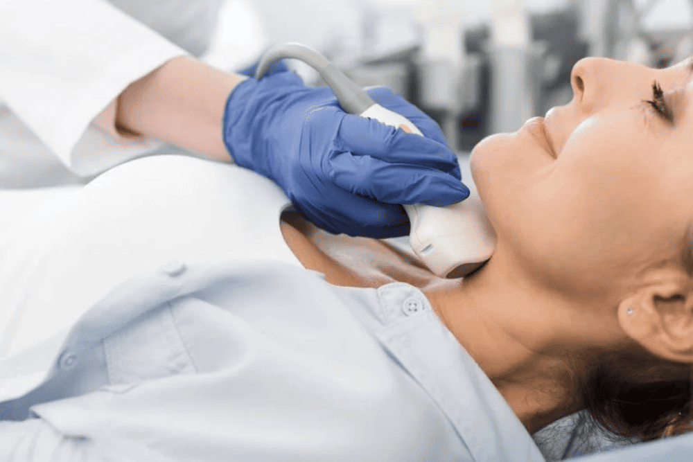
Finding a lump in your neck can raise many questions. Hearing the term “thyroid nodule” from your doctor can make those concerns feel more serious, especially when cancer is the first thing that comes to mind. The good news is that thyroid nodules are often benign. When a nodule is found, a thyroid ultrasound can provide important details about its size, shape, composition, and other features that may not be apparent from a physical exam alone.
C/V ENT Surgical Group provides thyroid and neck evaluation for patients in Encino who have a new or previously identified nodule. Ultrasound findings can help put the nodule in context, while the rest of the evaluation determines whether monitoring, additional testing, or other follow-up is appropriate.
What Is a Thyroid Ultrasound and Why Is It Used?
A thyroid ultrasound uses high-frequency sound waves to build a real-time image of the thyroid gland. A handheld device called a transducer sends sound waves into the neck. The waves bounce back differently depending on the tissue they strike. A computer turns these signals into a grayscale image. Your doctor studies this image as the scan happens.
This thyroid imaging method lets a clinician assess a nodule’s size and location. It also shows the nodule’s internal makeup and how its borders relate to the surrounding thyroid tissue. Because ultrasound provides detailed, real-time images without ionizing radiation, it is a primary imaging tool for evaluating thyroid nodules once a lump or abnormality is identified. A neck ultrasound can also evaluate nearby structures, including cervical lymph nodes. This makes it a practical starting point that gives your care team a broad view of your thyroid health without any incision or radiation.
Also Read: When to Worry About Thyroid Nodules?
What Can an Ultrasound Reveal About a Thyroid Nodule?
Once a nodule is found, ultrasound characterizes several distinct features. Clinicians record these features one by one, then read them together as a pattern.
Composition
The first trait a clinician checks is composition. This means whether the nodule is solid, cystic (fluid-filled), or mixed, with both solid and cystic parts. Composition matters because solid tissue and fluid-filled tissue carry different levels of clinical concern. This distinction often shapes how the rest of the report is read.
Echogenicity, Shape, and Margins
Several other ultrasound features of thyroid nodules are assessed together, including:
- Echogenicity, describing whether the nodule appears hypoechoic (darker), hyperechoic (brighter), or isoechoic (similar in tone) relative to the surrounding thyroid tissue
- Shape, including whether the nodule is wider than it is tall, or taller than it is wide
- Margins, meaning whether the nodule’s borders are smooth and well circumscribed or irregular and poorly defined
Calcifications and Size
Ultrasound can also identify echogenic foci, including certain types of calcifications within or around a nodule. These may be described as punctate echogenic foci, larger macrocalcifications, or peripheral rim calcifications, depending on their appearance. Size is measured in multiple dimensions at the time of the scan. Doctors often track them at later visits to check for growth.
A thyroid nodule can’t be fully assessed from one ultrasound feature alone. Your doctor considers the nodule’s size, shape, margins, composition, and other characteristics together when reviewing your ultrasound results. These findings are often listed in technical terms on the report, but each term refers to something the radiologist sees as a specific, observable trait. The combination of findings is what gives the report its clinical meaning.
Can an Ultrasound Tell if a Thyroid Nodule Is Cancerous?
Ultrasound alone cannot confirm or exclude cancer. Sonographic evaluation can flag features linked to a higher or lower chance of cancer. A nodule with irregular margins, a taller-than-wide shape, or certain calcification patterns may be seen as more suspicious. This may prompt further workup. A nodule with smooth margins and a mostly cystic makeup is often viewed as lower risk. This can still vary from case to case.
It also helps to know that imaging has limits. Some benign nodules can show features that look concerning on ultrasound. Some cancerous nodules don’t show every classically suspicious trait. Because of this, suspicious thyroid nodule imaging usually leads to more evaluation rather than an immediate conclusion. Your doctor may generally weigh the sonographic findings alongside your clinical history, family history, lab-based thyroid testing, and nodule size. This layered approach helps avoid both unnecessary intervention and missed findings that need closer attention.
How Is a Thyroid Nodule Evaluated After the Ultrasound?
After your thyroid nodule evaluation with ultrasound is complete, your doctor builds a management plan based on the full clinical picture.
Observation and Laboratory Testing
In many cases, a nodule with reassuring features is managed with surveillance. This means periodic repeat imaging to watch for any change. In other cases, your doctor may order lab studies, such as thyroid function tests, to check how well the gland is working.
Fine-Needle Aspiration Biopsy
If a nodule’s ultrasound appearance and size meet criteria for further evaluation, your doctor may recommend a fine-needle aspiration biopsy. Interval growth or changes in the nodule’s features may also affect follow-up decisions. During FNA, a thin needle, often guided by ultrasound, is used to collect cells from the nodule for laboratory examination. Not every thyroid nodule requires biopsy.
Also Read: Causes Of Thyroid Nodules and Its Treatment Options
What Happens During a Thyroid Ultrasound?
A thyroid ultrasound usually takes place while you are lying on your back. You may be asked to tilt your head back slightly to give the clinician better access to the front of your neck. The clinician applies a small amount of gel to your skin and gently moves an ultrasound transducer over the area to capture images of your thyroid from different angles.
The transducer uses sound waves to create images, so the exam doesn’t involve ionizing radiation. You may feel some light pressure as it moves across your neck, but the procedure is generally painless. The gel may feel cool when it is first applied. You may also be asked to swallow during the exam so the clinician can see how the thyroid moves and capture clearer images.
After the scan, the appropriate clinician or radiologist reviews the images. Your results may describe features such as the nodule’s size, composition, shape, and margins. Depending on these findings and your overall thyroid health, your doctor may recommend routine monitoring, additional testing, or specialist evaluation.
If you have questions about a thyroid nodule or your ultrasound results, discuss them with a qualified clinician who can interpret the findings in the context of your medical history.
When Should You Seek Evaluation for a Thyroid Nodule?
Many thyroid nodules cause no noticeable symptoms. They are often discovered by chance, either during a self-exam or when a clinician examines the neck for another reason. When symptoms do occur, they may include:
- A lump or swelling that you can see or feel in your neck
- Trouble swallowing or a sensation that something is pressing against your throat
- Changes in your voice, such as persistent hoarseness
- Ongoing pressure, tightness, or fullness in the neck
If you notice these symptoms, or if a thyroid nodule has already been found, an evaluation can help clarify what is happening. Early assessment doesn’t necessarily mean something serious is wrong. It gives your doctor an opportunity to review the nodule and determine whether monitoring, additional thyroid testing, or another type of evaluation is appropriate. The team at C/V ENT Surgical Group provides comprehensive ENT and thyroid evaluation for patients throughout the Encino area who want a clearer understanding of a new or existing thyroid mass.
Also Read: Thyroid Nodule Treatment: Thyroid Gland Evaluation and What to Expect
Conclusion
A thyroid ultrasound gives a detailed, noninvasive look at a nodule’s size, structure, and sonographic traits. This gives your doctor useful clinical information to work with. These features can raise or lower suspicion for cancer, but ultrasound alone can’t confirm or exclude thyroid cancer. It works best as one part of a broader diagnostic process. That process may include lab testing, surveillance imaging, or a biopsy when clinically needed.
If a thyroid nodule was recently found or your imaging results left you with questions, a qualified specialist can help you understand what the findings mean and what to consider next. C/V ENT Surgical Group is available to help Encino-area patients better understand their results and determine the right path forward for their thyroid health. Schedule your consultation at C/V ENT Surgical Group’s West Hills or Encino office today.
Frequently Asked Questions
What can an ultrasound reveal about a thyroid nodule?
It can characterize the nodule’s size, composition, shape, margins, echogenicity, and calcification pattern, all of which help a clinician assess the overall level of concern.
What is the difference between a benign and suspicious thyroid nodule on ultrasound?
Ultrasound features associated with lower suspicion may include a cystic or spongiform composition and other reassuring characteristics. Features that can raise suspicion include a solid or very hypoechoic appearance, irregular margins, a taller-than-wide shape, or certain echogenic foci. No single feature usually determines whether a nodule is cancerous, so clinicians interpret the overall ultrasound pattern along with nodule size and clinical history.
What happens after an ultrasound finds a thyroid nodule?
Depending on the findings, your doctor may recommend surveillance with follow-up imaging, additional laboratory testing, or a fine-needle aspiration biopsy.
Can a thyroid ultrasound determine if a nodule is cancerous?
Not on its own. It can identify features associated with higher or lower suspicion, but a definitive diagnosis typically requires further evaluation, including biopsy when indicated.
When should I see an ENT specialist about a thyroid nodule in Encino?
If you notice a neck lump, difficulty swallowing, voice changes, or persistent neck pressure, or if you simply want a thorough evaluation of a known nodule, reaching out to a specialist at C/V ENT Surgical Group is a reasonable next step.